TDP-43: A Potential Therapeutic Target for ALS
Introduction
When people think of the drug creation process, it is often thought that the identification of a therapeutic target would immediately lead to the creation of a new drug or therapy. However, in practice, this is rarely true because there are still many considerations before an effective treatment can be developed. This phenomenon is exemplified in the identification of a salt-bridge of the TAR DNA binding protein-43 (TDP-43) as a novel therapeutic target for the treatment of amyotrophic lateral sclerosis (ALS)1.
Amyotrophic Lateral Sclerosis
ALS is a motor neuron disease characterized by the breakdown and death of nerve cells in the brain, brainstem and spinal cord2. As nerve cells in both the brain and spinal cord are affected, the transmission of information between the two are interrupted, resulting in the loss of voluntary muscle movement2. It is estimated that approximately 2500 to 3000 Canadians over 18 currently live with ALS3. Early symptoms of the disease include muscle weakness, difficulty performing delicate tasks using the fingers or hands and uncoordinated movements. As ALS progresses, other symptoms include impairment of the tongue, mouth or voice box, stiffness in the legs, and uncontrolled muscles twitches4.
There are various types of ALS, differentiated by their symptoms and their genetic cause or lack thereof2. Research indicates that there are several types of hereditary ALS either through autosomal dominant or recessive methods of inheritance2. For example, an inherited form of ALS is known as ALS15. ALS1 is an autosomal dominant form of ALS due to changes in the superoxide dismutase-1 gene5. Only 5 to 10 percent of ALS patients have a family history of ALS and the majority of people with ALS have sporadic ALS, which occurs despite the lack of family history of ALS2.
Despite the extensive research, the exact underlying cause of the disease is unknown, although many mechanisms have been proposed. One mechanism involves TDP-43, a protein known to accumulate in the nerve cells of ALS patients that has the ability to cause nerve cell death1. Recently, researchers have identified a specific structure of TDP-43 that is the newly suspected cause of nerve cell death in ALS patients1. This discovery is exciting as there are currently no cures or effective treatments to halt or stop the progression of the disease, and the newly identified structure of the TDP-43 protein could serve as a potential therapeutic target.
Past Research
“This discovery is exciting as there are currently no cures or effective treatments to halt or stop the progression of the disease, and the newly identified structure of the TDP-43 protein could serve as a potential therapeutic target.”
Past research has established that TDP-43 plays various roles normally within the body as a DNA and RNA binding protein6. One of its main functions is to repress the transcription process in cells, especially those infected by HIV-16. Another function of the protein is to regulate alternative splicing of the cystic fibrosis transmembrane conductance regulator gene and Apolipoprotein A-II gene6. In spinal motor neurons, TDP-43 has also been shown to possibly regulate mRNA stability, transport and translation of neurons6,7.
In normal cells, TDP-43 is mostly present within the nucleus, although it can move between the nucleus and cytoplasm8. However, due to unknown reasons, it can cluster in the cytoplasm and deplete TDP-43 from the nucleus of the cell8. If this continues, the TDP-43 in the cytoplasm undergoes liquid-liquid phase separation, where the proteins condense into a dense phase, similar to liquid droplets, and becomes less mobile8,9. This results in a build-up of dense TDP-43 in the cytoplasm8. In cases such as these, the proteins are abnormal. For example, the TDP-43 is hyperphosphorylated, meaning that multiple sites on the protein contain phosphate groups which alter its normal function7,8. Alternatively, the TDP-43 are ubiquitinated, with ubiquitin molecules attached to the protein10. Around 97% of ALS cases involve the build-up of TDP-43 and many mutations within the gene that codes for the TDP-43 protein serve as a cause of hereditary ALS8.

Figure 1. A depiction of how TDP-43 accumulation in the cytoplasm of a cell (as indicated by the orange spots) lead to degeneration of a
motor neuron.
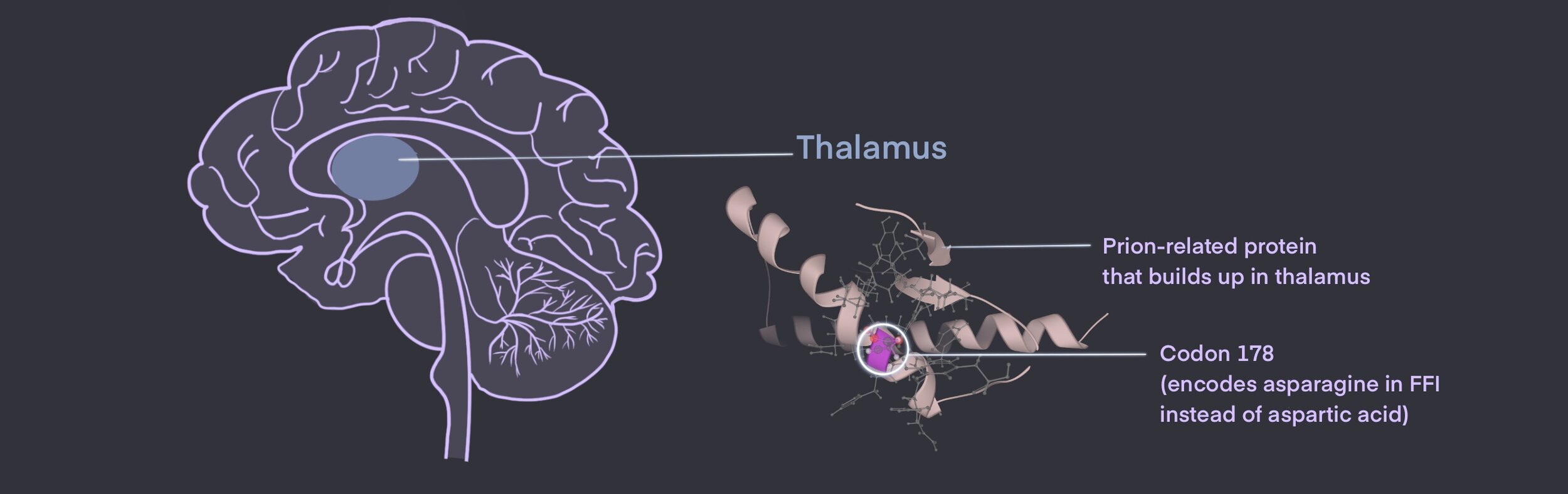
Structurally, it is known that TDP-43 has 2 components which serve as RNA recognition sites: RRM1 and RRM212. Studies with fruit flies have shown that when the RRM1 component is deleted, RNA binding is prevented and the toxic build-up of TDP-43 is eliminated12. Studies have also shown that the stability of TDP-43 is important in determining whether it would be toxic to the cell or not1. Therefore, researchers suspected that RNA binding and TDP-43 stability was an important part of ALS, however, they had little information until now.
“Without RNA binding, TDP-43 was quickly degraded and TDP-43 mediated toxicity completely mitigated1. Therefore, it was clear that if excess TDP-43 proteins are degraded through disruptions to the TDP-43 structure, nerve cells would have a chance of survival.”
New Research
To investigate the influence of TDP-43 on ALS, researchers manipulated the structure of TDP-43. In doing so, they were able to identify a specific part of the protein that causes nerve cell death. Between the RRM1 and RRM2 recognition sites, there is a salt bridge1. This salt bridge is important not only for RNA binding but also for maintaining the stability of the protein1. TDP-43 proteins that lack this salt bridge cannot bind RNA, and are incapable of causing the degradation of nerve cells1. To explore the question of whether the structure of this salt bridge could be altered to cause a change in TDP-43 toxicity, mutations were introduced to the RRM1-RRM2 salt bridge1. Then, automated microscopy, a technique where images from a microscope are taken overtime and analyzed using a computer, was used to determine the rate at which nerve cells die1. Through this method, imaging showed that the modified TDP-43 protein, with a disrupted salt bridge, was less toxic than the non-modified one1. Specifically, researchers found that if they introduced any mutations to the arginine amino acid present at the R151 residue (a part of the salt bridge), RNA binding was prevented1. Without RNA binding, TDP-43 was quickly degraded and TDP-43 mediated toxicity completely mitigated1. Therefore, it was clear that if excess TDP-43 proteins are degraded through disruptions to the TDP-43 structure, nerve cells would have a chance of survival1.
Future Directions
“ If it can be confirmed that TDP-43 build-up is present in every, or most ALS, patients, then a future therapy disrupting the salt-bridge may be used to treat or cure all types of ALS, despite their different causes.”
Knowing that it is the structure of TDP-43 which causes nerve cell death opens the door to developing new therapies for ALS. For example, a new drug could target the arginine amino acid of the R151 residue to disrupt the salt bridge. This way, extra TDP-43 proteins would be degraded and nerve cell death could be prevented. However, although a new target has been identified, this does not mean that the development of a new drug is imminent. Since the TDP-43 protein is essential for normal cell function, completely disrupting the structure of all TDP-43 proteins or knocking out the gene that codes for TDP-43 would cause major disruptions in normal life processes. Therefore, the question is, how do we create a drug that promotes the degradation of excess TDP-43 while ensuring that normal TDP-43 functioning is not disrupted? Researchers have yet to answer this question. This shows that the process of developing new therapies is extremely complex and has to take into account a variety of factors in addition to therapeutic targets.
Nonetheless, this research is especially interesting considering the high level of ALS cases (97%) which involve the build-up of TDP-438. If it can be confirmed that TDP-43 build-up is present in every, or most ALS, patients, then a future therapy disrupting the salt-bridge may be used to treat or cure all types of ALS, despite their different causes. This would make the management of ALS easier for doctors as well as their patients. Even if a therapy is not developed from this research, the process of researching TDP-43 brings us one step closer to elucidating the underlying mechanisms behind ALS and in determining future directions for ALS drug research.
Ilziba Yusup
References
1. Flores BN, Li X, Malik AM, Martinez J, Beg AA, Barmada SJ. An intramolecular salt bridge linking TDP43 RNA binding, protein stability, and TDP43-dependent neurodegeneration. Cell Reports. 2019;27(4):1133-1150.e8. doi:10.1016/j.celrep.2019.03.093
2. Amyotrophic Lateral Sclerosis. NORD (National Organization for Rare Disorders). Accessed March 10, 2021. https://rarediseases.org/rare-diseases/amyotrophic-lateral-sclerosis/
3. What is ALS? ALS Society of Canada. Accessed March 10, 2021. https://www.als.ca/what-is-als/
4. Amyotrophic Lateral Sclerosis (ALS) Fact Sheet | National Institute of Neurological Disorders and Stroke. Accessed March 10, 2021. https://www.ninds.nih.gov/Disorders/Patient-Caregiver-Education/Fact-Sheets/Amyotrophic-Lateral-Sclerosis-ALS-Fact-Sheet
5. Andersen PM, Borasio GD, Dengler R, et al. EFNS task force on management of amyotrophic lateral sclerosis: guidelines for diagnosing and clinical care of patients and relatives. An evidence-based review with good practice points. Eur J Neurol. 2005;12(12):921-938. doi:10.1111/j.1468-1331.2005.01351.x
6. Mackenzie IRA, Rademakers R. The role of TDP-43 in amyotrophic lateral sclerosis and frontotemporal dementia. Curr Opin Neurol. 2008;21(6):693-700. doi:10.1097/WCO.0b013e3283168d1d
7. Fallini C, Bassell GJ, Rossoll W. The ALS disease protein TDP-43 is actively transported in motor neuron axons and regulates axon outgrowth. Hum Mol Genet. 2012;21(16):3703-3718. doi:10.1093/hmg/dds205
8. Loganathan S, Lehmkuhl EM, Eck RJ, Zarnescu DC. To be or not to be…toxic—Is RNA association with TDP-43 complexes deleterious or protective in neurodegeneration? Front Mol Biosci. 2020;6. doi:10.3389/fmolb.2019.00154
9. Alberti S, Gladfelter A, Mittag T. Considerations and challenges in studying liquid-liquid phase separation and biomolecular condensates. Cell. 2019;176(3):419-434. doi:10.1016/j.cell.2018.12.035
10. Dong Y, Chen Y. The role of ubiquitinated TDP-43 in amyotrophic lateral sclerosis. Neuroimmunology and Neuroinflammation. 2018;5. doi:10.20517/2347-8659.2017.47
11. Broeck LV, Callaerts P, Dermaut B. The TDP-43 Gain- versus Loss-of-Function Debate.; 2013.https://www.cell.com/trends/molecular-medicine/fulltext/S1471-4914(13)00196-2#fig0005.
12. Ayala YM, Pantano S, D’Ambrogio A, et al. Human, Drosophila, and C.elegans TDP43: nucleic acid binding properties and splicing regulatory function. J Mol Biol. 2005;348(3):575-588. doi:10.1016/j.jmb.2005.02.038
Cite This Article:
Yusuo I., Rashid S. & Wong V. TDP-43: A Potential Therapeutic Target for ALS. Illustrated by S. Montakhaby. Rare Disease Review. October 2021.